Fabulous How To Read A Breast Ultrasound Report Write Good Methodology For Research
The Radiology Assistant Ultrasound Of The Breast
An ultrasound image of the same lesion suggests that the lesion is solid. The anterior margin of the tear is adjacent to. The upper grey layer is the skin. Your breasts look the same they are. It is used to help diagnose breast lumps or other abnormalities found during a physical exam or on a mammogram or breast MRI. Ultrasound is safe noninvasive and does not use radiation. Ultrasound is also used to diagnose problems such as complications from mastitis an infection that occurs most often during breast-feeding assessing abnormal nipple discharge or problems with breast implants. The X-ray image below shows a suspect breast mass of about 1 cm in diameter. 8 rows Theres no significant abnormality to report. What more it is even possible to predict the age and the gender of the baby early on in pregnancy something which was impossible a few years ago.
LOL The findings were.
Breast ultrasound is performed very commonly after a mammogram to tell whether a lump in a breast is a cyst a fluid-filled sac or a solid mass which might be breast cancer. Pathology reports contain information about the patient the tissue specimen being evaluated and the final diagnosis. An ultrasound image of the same lesion suggests that the lesion is solid. Shoulder pain evaluate for rotator cuff abnormality Findings. Breast cancers because many new blood vessels to develop in these new blood vessels will develop and surround the breast cancer. Some architectural distortion is also apparent.
The procedure is straightforward. Through a lot a hard-work I finally got the report sent to my PCP. Breast cancer is among the most common causes of cancer deaths today coming fifth after lung stomach liver and colon cancers. During my mammo a tumor was foundI was asked to wait same day for a ultrasoundThe ultrasound was just to make sure what it could beCyst tumor or maybe nothingI had the ultrasound that same dayDoctor read the Xray and said it was a tumor and he didnt think it was anything BUT I must have a biopsyAfter the biopsy is scheduled you usually know in a couple days. Pathology reports contain information about the patient the tissue specimen being evaluated and the final diagnosis. Therefore the MRI can see these blood vessels around the breast cancer and you actually can see the breast cancer. Breast ultrasound is performed very commonly after a mammogram to tell whether a lump in a breast is a cyst a fluid-filled sac or a solid mass which might be breast cancer. Its most important use however is to determine anomalies andgenetic disorders in the foetus early on in pregnancy that gives time for the doctors and the expectant mother to think about corrective measures even while the baby is in the womb. 1to get a data for accuracy of musculoskeletal Ultrasound 2to determine accuracy of MSK for different jointsmusclesligaments and tendons. Figure 5-1 Normal breast ultrasound images.
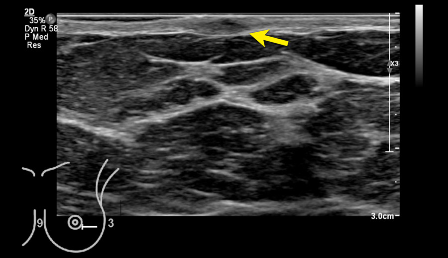
To read an ultrasound picture look for white spots on the image to see solid tissues like bones and dark spots on the image to see fluid-filled tissues like the amniotic fluid in the uterus. The anterior margin of the tear is adjacent to. A and B Normal breast ultrasound scans in fatty mixed and dense breasts. This example demonstrates a report on multiple breast imaging procedures. If the cyst is not entirely clear on the ultrasound a doctor may drain it with a needle and send the fluid for testing to check for cancer cells. Each patients cancer. Figure 5-1 Normal breast ultrasound images. Targeted sonography was performed of the palpable area of the right breast at 12 oclock that was diagramed on the requision for this study. Press Command to enlarge images - Scroll for text. So most likely youll get.
Pathology reports contain information about the patient the tissue specimen being evaluated and the final diagnosis. The sensitivity of mammography for breast cancer detection in women over 50 years is well over 80 and in the symptomatic population when combined with breast ultrasound US this figure increases to around 90 It might then be asked why another imaging modality such as breast. Your breasts look the same they are. Dark fatty lobules separated by sharp thin Cooper ligaments. An ultrasound image of the same lesion suggests that the lesion is solid. Unlabeled and labeled ultrasounds of a normal fatty breast show the thin white superficial skin line. Breast cancer is among the most common causes of cancer deaths today coming fifth after lung stomach liver and colon cancers. If breast cancer spreads it often goes first to the nearby lymph nodes under the arm called axillary lymph nodes. If any of your underarm lymph nodes were enlarged found either by physical exam or with an imaging test like ultrasound or mammogram they may be biopsied at the same time as your breast. Following a screening mammogram the patient was asked to return for additional imaging and an ultrasound on the breast for further evaluation of a mammographic mass.
It can help your healthcare provider find breast problems. Ultrasound is safe noninvasive and does not use radiation. Each patients cancer. Hi everyone I posted a couple of days ago while waiting for my results of my ultrasound. Theyll move the transducer. ULTRASOUND be listed by breast by location within in the breast and by size. Shoulder pain evaluate for rotator cuff abnormality Findings. Targeted sonography was performed of the palpable area of the right breast at 12 oclock that was diagramed on the requision for this study. If youre 12 weeks along in the pregnancy you may be able to make out your babys head and if youre 20 weeks along you may even see the spine heart feet and eyes. March 11 2017 Patient Name.
LOL The findings were. Breast cancer is among the most common causes of cancer deaths today coming fifth after lung stomach liver and colon cancers. It is used to help diagnose breast lumps or other abnormalities found during a physical exam or on a mammogram or breast MRI. Chazz Michael Michaels Registration Number. Pathologists evaluate the tissue within a breast core biopsy or surgical specimen to diagnose the type of cancer as well as evaluate the presence of various prognostic factors. It is the most common cause of cancer death in women In 2005 alone 519 000 deaths were recorded due to breast cancer This means that one in every 100 deaths worldwide and almost one in every 15 cancer deaths were due to breast cancer. The procedure is straightforward. On your report the radiologist may describe enhancement or may describe an enhancing mass. There is a focal anechoic tear of the anterior distal aspect of the supraspinatus tendon measuring 1 cm short axis by 15 cm long axis. On the day of scheduled breast ultrasound wear a two piece outfit.